Your eye – How you see
Understanding glaucoma requires a basic understanding of the eye, its various parts, and how it works.
The eye captures visual information about the world and sends it in the form of nerve impulses to the brain. The brain processes this information into the “pictures” we see.
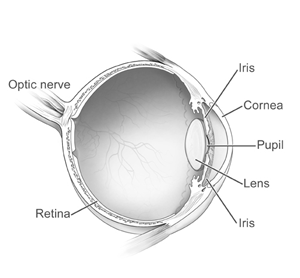
The outer, white layer of the eyeball is the sclera, a tough, leathery protective shell.
The front, transparent portion of the shell is the cornea, through which light enters the eye. The cornea is much like the lens of a camera, providing the eye with much of its focusing power.
The coloured portion of the eye is the iris. The iris contains muscles that control the size of the pupil. This regulates the amount of light entering the eye. The pupil constricts in bright light and dilates in dim light, adjusting the amount of light that passes through to the retina. The difference between blue and brown irises is the amount of pigment in the front portion of the iris.
The anterior chamber, or front compartment of the eye, is filled with a clear watery fluid called aqueous. Aqueous is continuously made by the ciliary body, a tiny gland behind the iris. Aqueous provides the structures of the eye with oxygen and vital nutrients. The aqueous is also responsible for the pressure (IOP) inside the eye. This pressure is necessary to maintain the shape of the eye.
Aqueous fluid then leaves the anterior chamber at the open angle where the cornea and iris meet. When the fluid reaches the angle, it flows through a spongy meshwork, like a drain, and leaves the eye. The fluid then drains back into the bloodstream. Normally, the amount of fluid produced is balanced by the amount draining away, so the pressure in the eye stays constant. In glaucoma the fluid drains too slowly out of the eye, fluid builds up and pressure rises.
The lens behind the iris is transparent like the cornea. The lens adjusts its shape and thickness to focus the image onto the retina. When we read, the eye accommodates to refocus a near image. The ability to accommodate decreases steadily throughout life. Presbyopia occurs when there is not enough accommodative power remaining to read without glasses, usually in the early 40s.
After passing through the lens, the light reaches the retina. The retina then delivers electrical signals to the optic nerve.
The optic nerve is made up of over one million nerve cells. The optic nerve carries visual information to the brain. The brain processes these signals into a “picture”, or visual image. In glaucoma damage to the optic nerve results in loss of vision.
What is glaucoma